Welcome to the Amira-Avizo Software Use Case Gallery
Below you will find a collection of use cases of our 3D data visualization and analysis software. These use cases include scientific publications, articles, papers, posters, presentations or even videos that show how Amira-Avizo Software is used to address various scientific and industrial research topics.
Use the Domain selector to filter by main application area, and use the Search box to enter keywords related to specific topics you are interested in.
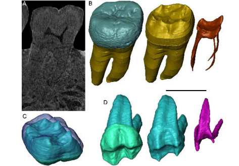
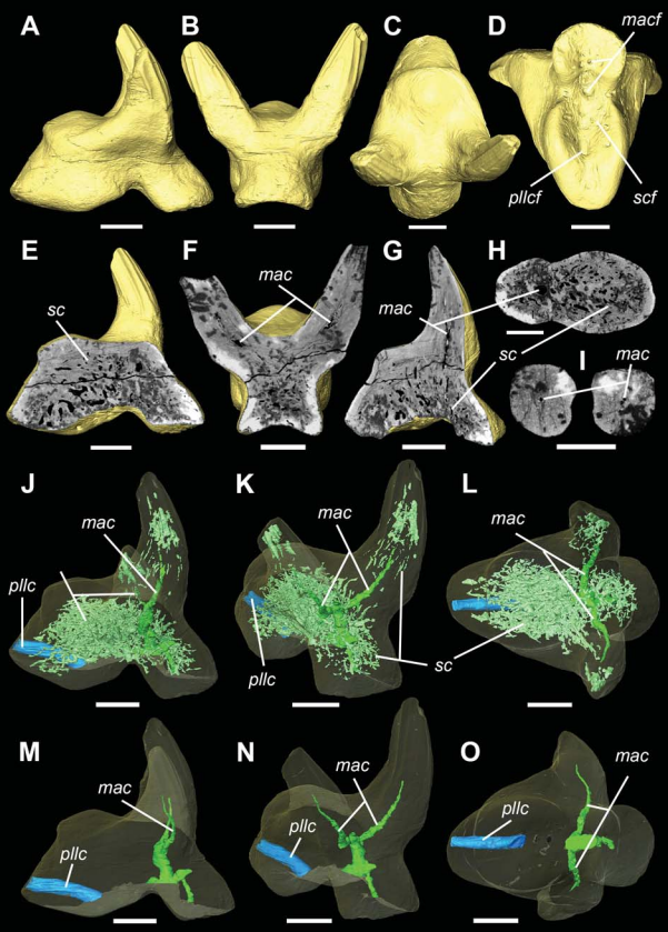
Exploring hominin and non-hominin primate dental fossil remains with neutron microtomography
Fossil dental remains are an archive of unique information for paleobiological studies. Computed microtomography based on Xray microfocus sources (X-µCT) and Synchrotron Radiation (SR-µCT) allow subtle quantification at the micron and sub-micron scale of the meso- and microstructural signature imprinted in the mineralized tissues, such as enamel and dentine, through highresolution “virtual histology”. Nonetheless, depending on the degree of alterations undergone during fossiliza... Read more
Clément Zanolli, Laboratory AMIS, UMR 5288, University of Toulouse III - Paul Sabatier, France, and al.
Endosseous oral implant is applied for orthodontic anchorage in subjects with multiple tooth agenesis. Its effectiveness under orthodontic loading has been demonstrated clinically and experimentally. This study investigates the deformation and stress on the bone and implant for different bite forces by three-dimensional (3D) finite element (FE) methods. A numerical simulation of deformation and stress distributions around implants was used to estimate the survival life for implants. The model... Read more
Hsin-Chung Cheng, Boe-Yu Peng, May-Show Chen, Chiung-Fang Huang, Yi Lin, and Yung-Kang Shen
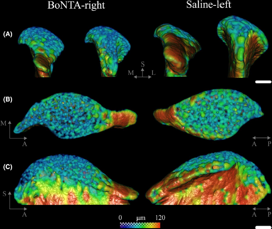
Masseter muscle function influences mandibular bone homeostasis. As previously reported, bone resorption markers increased in the mouse mandibular condyle two days after masseter paralysis induced with botulinum toxin type A (BoNTA), followed by local bone loss.
This study aimed to evaluate the bone quality of both the mandibular condyle and alveolar process in the mandible of adult mice during the early stage of a BoNTA‐induced masseter muscle atrophy, using a combined 3D histomorpho... Read more
Julián Balanta‐Melo, María Angélica Torres‐Quintana, Maximilian Bemmann, Carolina Vega, Constanza González, Kornelius Kupczik, Viviana Toro‐Ibacache, Sonja Buvinic
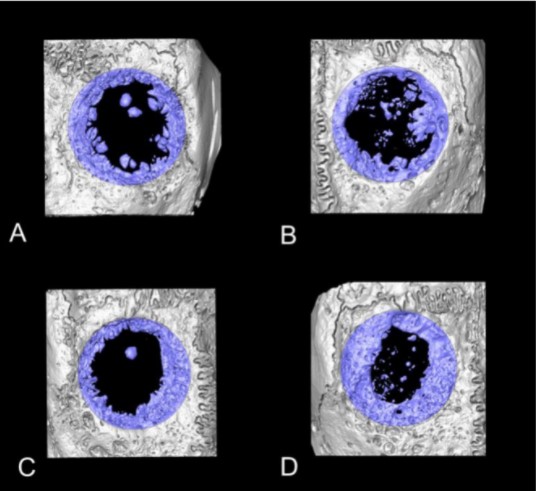
Collagen-Based Matrices for Osteoconduction: A Preclinical In Vivo Study
The aim of this study was to evaluate the influence of additional hydroxyapatite (HA) in collagen-based matrices (CM) and membrane placement on bone formation in calvarial defects.
Critical size defects in the calvaria of 16 New Zealand White Rabbits were randomly treated with CM or mineralized collagen-based matrices (mCM). Half of the sites were covered with a collagen membrane. Animals were euthanized after 12 weeks of healing. The samples were studied by micro-CT and histology. New... Read more
Hiroki Katagiri, Yacine El Tawil, Niklaus P. Lang, Jean-Claude Imber, Anton Sculean, Masako Fujioka-Kobayashi and Nikola Saulacic
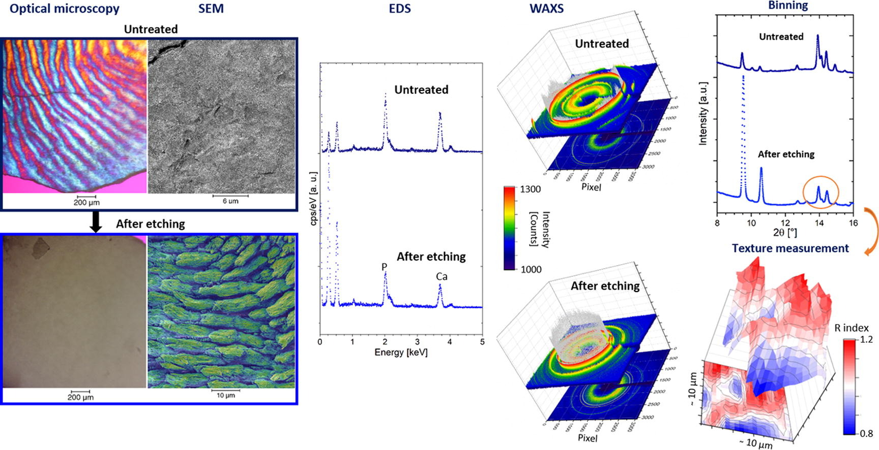
Enamel caries is a highly prevalent worldwide disease that involves the demineralisation of the outer tooth structure. In this study, we report the analysis of artificially demineralised human enamel sections (‘slices’) etched using lactic acid (2% v/v) in comparison with healthy enamel using correlative techniques of optical and electron microscopy, as well as scanning diffraction. Demineralisation of the enamel was characterised at the micron to sub-micron scale. The structure of the he... Read more
Cyril Besnard, Robert A. Harper, Thomas E. J. Moxham, Jonathan D. James, Malte Storm, Enrico Salvati, Gabriel Landini, Richard M. Shelton, Alexander M.Korsunsky
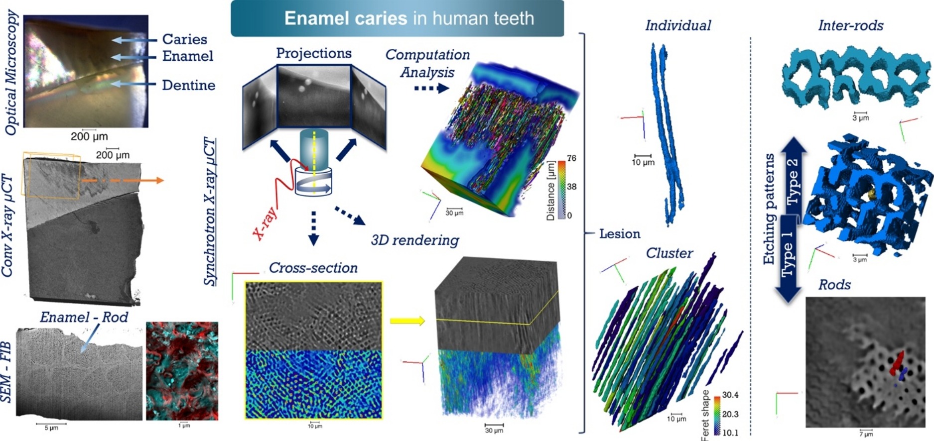
Unprecedented combination of resolution, field of view and contrast for the analysis human enamel carious lesions was achieved. Synchrotron X-ray micro-computed tomography revealed sub-micron details of enamel rod and inter-rod regions inaccessible by laboratory tomography. Successful segmentation and labelling allowed the extraction of enamel etching patterns and statistics. Correlation was obtained between synchrotron X-ray micro-tomography and FIB-SEM cross-sec... Read more
Cyril Besnard, Robert A. Harper, Thomas E. J. Moxham, Jonathan D. James, Malte Storm, Enrico Salvati, Gabriel Landini, Richard M. Shelton, Alexander M.Korsunsky
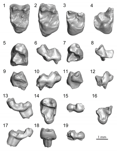
Variation of 3D outer and inner crown morphology in modern human mandibular premolars
This study explores the outer and inner crown of lower third and fourth premolars (P3, P4) by analyzing the morphological variation among diverse modern human groups.
We studied three‐dimensional models of the outer enamel surface and the enamel–dentine junction (EDJ) from μCT datasets of 77 recent humans using both an assessment of seven nonmetric traits and a standard geometric morphometric (GM) analysis. For the latter, the dental crown was represented by ... Read more
Viktoria A. Krenn, Cinzia Fornai, Lisa Wurm, Fred L. Bookstein, Martin Haeusler, Gerhard W. Weber
During the acts of biting and chewing, the muscles of the jaw (consisting of the masseter, temporalis, and medial pterygoid in the elevator group, and lateral pterygoid as the main depressor) generate forces that dictate jaw kinematics . The movement of jaws hinges about the temporomandibular joint and are brought together by the muscles attached to respective bones through bone-tendon interfaces known as entheses . Thus, chewing forces affect aspects of craniofacial structure as well as bo... Read more
Kyle H.-Y. Chan, fourth-year undergraduate student in Molecular and Cell Biology, and Public Health at UC Berkeley, FeiFei Yang, Ph.D., postdoctoral scholar in the Laboratory of Multiscale Biomechanics and Biomineralization, School of Dentistry, UCSF, and Sunita P. Ho, Ph.D., Division of Biomaterials and Bioengineering, Department of Preventive and Restorative Dental Sciences, School of Dentistry, University of California San Francisco
Vascular structure of the earliest shark teeth
Here we use synchrotron tomography to characterise dental vasculature in the oldest known tooth-bearing sharks, Leonodus carlsi Mader, 1986 and Celtiberina maderi Wang, 1993. Three dimensional reconstruction of the vascular system and microstructure of both taxa revealed a complex and dense network of canals, including horizontal, ascending and secondary bifurcated canals, as well as histological features consistent with an osteodont histotype. However, L. carlsi and C. maderi also exhibit si... Read more
Carlos Martinez-Perez, Alba Martin-Lazaro, Humberto G Ferron, Martina Kirstein, Philip C.J. Donoghue, Hector Botella
Nonuniformity in ligaments is a structural strategy for optimizing functionality
Ligaments serve as compliant connectors between hard tissues. In that role, they function under various load regimes and directions. The 3D structure of ligaments is considered to form as a uniform entity that changes due to function. The periodontal ligament (PDL) connects the tooth to the bone and sustains different types of loads in various directions. Using the PDL as a model, employing a fabricated motorized setup in a microCT, we demonstrate that the fibrous network structure with... Read more
Gili R. S. Naveh, Jonathan E. Foster, Tomas M. Silva Santisteban, Xianrui Yang, and Bjorn R. Olsen
Paromomyidae has been thought to represent the longest-lived group of stem primates (plesiadapiforms), extending from the early Paleocene to late Eocene. We analyzed primate material from the late-middle Eocene of southern California that had initially been ascribed to cf. Phenacolemur shifrae. This material falls at the lowest end of the size range for the family. The Californian specimens also exhibit several dental features that are atypical for paromomyids, such as a strong paraconid on t... Read more
Sergi López-Torres, Mary T. Silcox, and Patricia A. Holroyd
Mechanical adaptation of trabecular bone morphology in the mammalian mandible
Alveolar bone, together with the underlying trabecular bone, fulfils an important role in providing structural support against masticatory forces. Diseases such as osteoporosis or periodontitis cause alveolar bone resorption which weakens this structural support and is a major cause of tooth loss. However, the functional relationship between alveolar bone remodelling within the molar region and masticatory forces is not well understood. This study investigated this relationship by comparing m... Read more
Peter J. Watson, Laura C. Fitton, Carlo Meloro, Michael J. Fagan, Flora Gröning
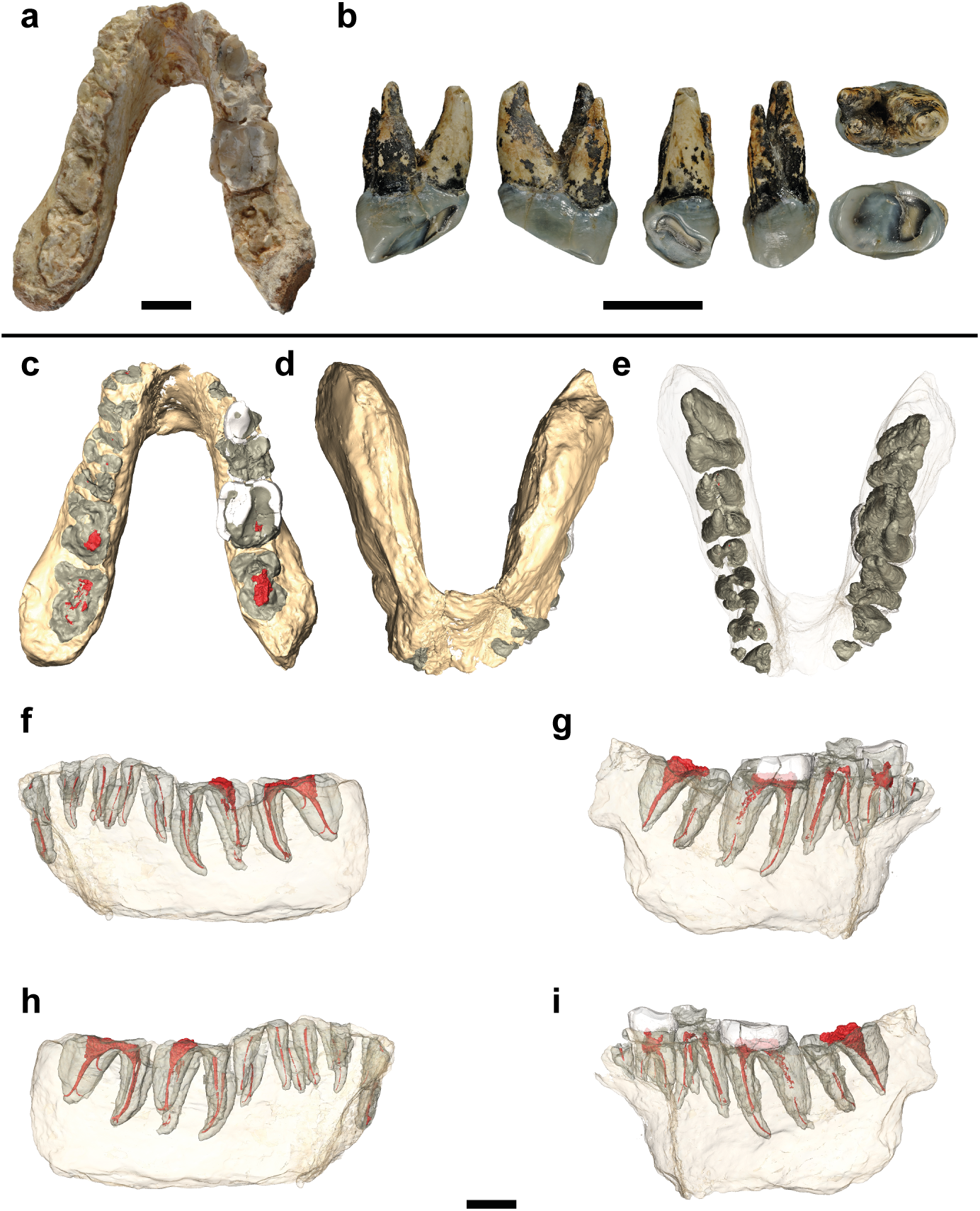
Potential hominin affinities of Graecopithecus from the Late Miocene of Europe
The split of our own clade from the Panini is undocumented in the fossil record. To fill this gap we investigated the dentognathic morphology of Graecopithecus freybergi from Pyrgos Vassilissis (Greece) and cf. Graecopithecus sp. from Azmaka (Bulgaria), using new μCT and 3D reconstructions of the two known specimens. Pyrgos Vassilissis and Azmaka are currently dated to the early Messinian at 7.175 Ma and 7.24 Ma. Mainly based on its external preservation and the previou... Read more
Jochen Fuss, Nikolai Spassov, David R. Begun, Madelaine Böhme
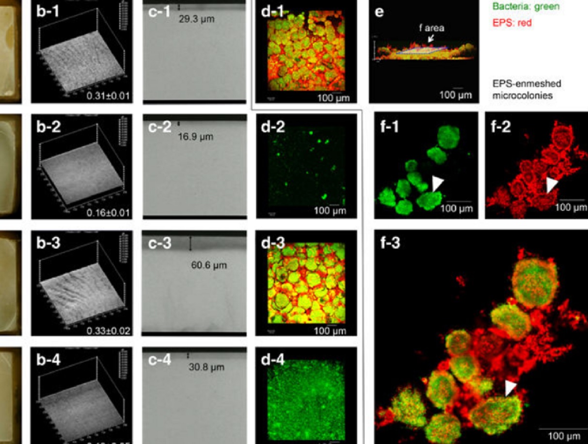
Acidic microenvironments created by bacterial clusters thriving in a polysaccharide matrix could be behind localized tooth decay. Jin Xiao of the University of Rochester Medical Center and Geelsu Hwang of the University of Pennsylvania with colleagues in the US mapped acidity changes across tooth enamel caused by the microstructure of dental plaque: a film of bacteria and the polysaccharide matrix they secrete. Using fluorescence microscopy, they studied the 3D architecture of plaque that for... Read more
Jin Xiao, Anderson T Hara, Dongyeop Kim, Domenick T Zero, Hyun Koo et al.